If a picture paints a thousand words, a CT scan paints 1 million.
In years past, surgeons would commonly conduct exploratory surgery—visually scouting within the patient to diagnose an ailment that a picture, in this case an X-ray, could not detect. While exploratory surgery still has its place in some applications, today complex ailments and the most life-threatening diseases are assessed with the aid of advanced medical imaging. Medical imaging is also used for basic scientific research, and to guide development and testing of new pharmaceuticals.
In spite of its high level of sophistication, medical imaging continues to improve at a dramatic pace, and IIT’s Medical Imaging Research Center is at the cutting edge of these technological innovations.
As Miles Wernick, Motorola Professor and director of MIRC explains, the group focuses on three broad areas of research—creating better images through development of new imaging devices and procedures, improving existing images through new mathematical approaches, and understanding the images that are obtained through the application of advanced computer algorithms. “Something most people don’t appreciate,” Wernick says, “is that modern medical images are not like photographs.” Rather than being captured directly, as images are in a digital camera, medical images (such as MRI) are computed by a complicated mathematical algorithm. “It’s in that computation that the image quality is largely defined,” he adds.
While many research teams specialize in one imaging modality or concentrate on the diagnosis of one type of disease, MIRC is unique in its breadth of areas explored, drawing on $6.3 million in recent research grants, mainly from several different components of the National Institutes of Health. In addition to refining existing imaging methods, MIRC investigators are exploring entirely new methods, such as thermoacoustic imaging, as well as sophisticated search engines and computerized analysis techniques for accessing and making sense of medical images.
Although IIT lacks a medical school of its own, MIRC—a branch of IIT’s Pritzker Institute of Biomedical Science and Engineering—benefits from close collaborations with the nearby University of Chicago Medical Center and Rush University Medical Center, as well as hospitals and medical schools elsewhere in the United States and in Canada.
The following are a few of the promising research areas currently under exploration at MIRC.
A Search Engine for Mammograms
Yongyi Yang (M.S. AMAT ’92, Ph.D. EE ’94), professor of electrical and computer engineering, is the principal investigator for an NIH-sponsored project to develop an innovative image-retrieval system—a sort of Google for mammography that conducts searches based on images rather than keywords. Given a patient’s mammogram, the software fetches and displays several similar mammograms from a vast library of stored images, providing the radiologist with examples of relevant past cases for comparison. “It’s like a doctor with a really big brain,” Yang says, referring to the search engine’s instant recall from tens of thousands of historical records. Should the search engine pull up images closely resembling those of the mammogram under consideration, it can inform the physician about past patient outcomes and offer useful hunches as to the benign or malignant nature of irregularities appearing in the patient at hand. The system could greatly enhance the usefulness of mammograms, which are notoriously tricky to interpret, and may also limit unnecessary biopsies and missed cancers. The project will be tested using the University of Chicago’s Hospital Information System, a database containing a large number of patient records.
Images on the Go
While brain imaging is typically accomplished by big, expensive machines like CT or MRI scanners, simpler, more portable devices are also being explored. Mark Anastasio (EE ’92), associate professor of biomedical engineering, along with colleagues at Washington University in St. Louis, is working on thermoacoustic imaging—a new technique that may be applied clinically within the next few years. The hybrid method combines electromagnetic waves with acoustical properties to deliver 3-D images. A short electromagnetic pulse is used to irradiate biological tissue, which absorbs this pulse, causing emission of acoustic signals that can be measured and used in conjunction with a mathematical algorithm to form a 3-D map of the tissue’s properties.
One of the significant advantages of thermoacoustic imaging is that it can be achieved using a compact device that can be wheeled into a hospital room, eliminating the need to transport patients to a separate imaging facility. It could also be used in ambulances, on battlefields, or in other remote settings. Further, thermoacoustic imaging provides an attractive alternative to X-ray imaging, replacing harmful ionizing radiation with safe microwave frequencies capable of penetrating the skull. Anastasio is working to perfect the mathematical tools, including image-reconstruction algorithms, needed to produce accurate images from this exotic form of data.
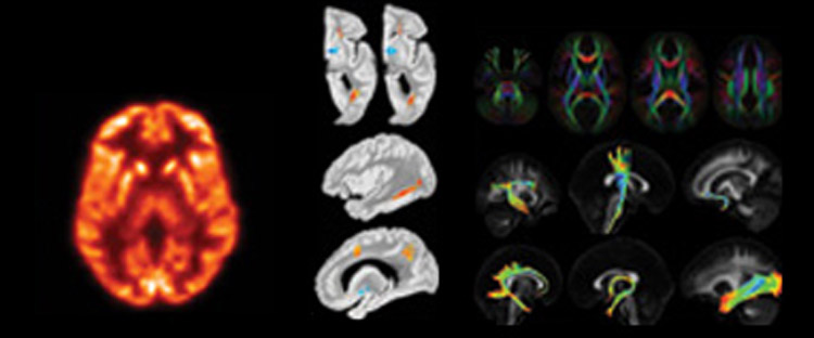
[Left to right]
- PET image of a brain showing glucose metabolism
- Image showing regions of iron accumulation (blue) and tissue degeneration (orange/red) in Alzheimer’s disease
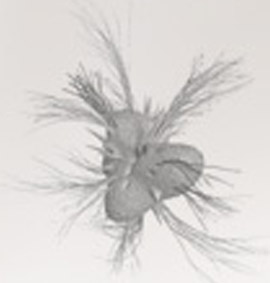
- Diffusion tensor imaging (DTI) nerve fibers
Mapping the Brain’s Superhighways
While the term gray matter—referring to concentrations of neuronal cell bodies in the brain—is familiar to most, the brain’s white matter is equally important. White-matter nerve fibers are highways of the central nervous system that connect different brain regions and are a focus of research for Konstantinos Arfanakis, BME associate professor. He has created an atlas of white-matter nerve fibers based on an MRI technique called diffusion tensor imaging (DTI). The atlas is now available to the scientific community: to date, more than 30 groups in more than 12 countries around the world are using the IIT atlas.
DTI is able to map neural tracts by measuring the amount of diffusion of water molecules within white-matter tissue. Within a healthy nerve fiber, water is mainly able to diffuse along the direction of nerve fiber tracts; therefore, by tracing the direction of highest diffusion, one can make a roadmap of the tracts.
When axons have been affected by disease, this diffusion property changes and provides clues to the onset of disease and its nature. Arfanakis is using DTI in a large-scale study of Alzheimer’s disease in collaboration with Rush Alzheimer’s Disease Center in Chicago to investigate associated microscopic changes in white-matter tissue.
Preliminary findings show that DTI may reveal changes in brain tissue long before a subject is diagnosed with Alzheimer’s disease. Early diagnosis of the disease will allow identification of the most appropriate human subjects to participate in tests of new drugs for Alzheimer’s patients. Also, early diagnosis of the disease will allow early intervention, which could minimize neuronal damage and associated impairment of mental function. And because conventional DTI techniques yield poor image quality in the brain regions that play a major role in the early stages of Alzheimer’s disease, Arfanakis is developing new DTI techniques to improve imaging in these regions.
BODY IMAGE
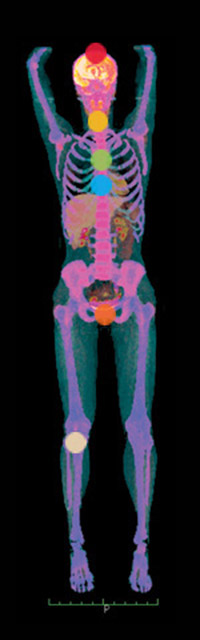
Imaging research at IIT’s Medical Imaging Research Center touches all areas of the body.
HEAD
MIRC is developing thermoacoustic imaging, a new technique allowing for a highly mobile imaging platform, combining sound and electromagnetic waves to produce a detailed, non-invasive image of the brain.
Diffusion tensor imaging, or DTI, has been applied to the detailed study of the brain’s white-matter fiber tracts. By examining differences in water diffusion through neural fibers, researchers hope to dramatically improve early diagnosis of Alzheimer’s disease and other degenerative brain ailments.
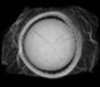
EYE
Image of the eye taken with a new X-ray setup at IIT
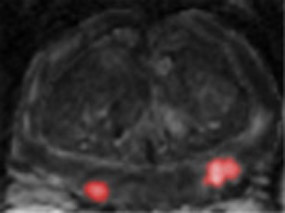
NECK
Identifying arterial plaques in the carotid artery through phase contrast imaging will help physicians identify plaques and determine which ones are in danger of rupturing.
BREAST
Microcalcifications in breast tissue and other abnormalities suggestive of disease are being examined through MIRC’s innovative image retrieval system—a kind of search engine that combs historical patient records, providing physicians with a wealth of diagnostically relevant information.
Phase contrast imaging and multiple-image radiography, two advanced forms of X-ray imaging, are being refined to provide more detailed and useful mammography images.
HEART
MIRC is developing new 4-D and 5-D approaches to imaging with single photon emission computed tomography (SPECT) that use advanced image-processing methods to permit new ways of imaging patients with heart disease.
MIRC is also developing image analysis methods based on CT scans, which identify vulnerable plaques in coronary arteries.
JOINTS, KNEES, ANKLES, WRISTS, ELBOWS,
FINGERS, TOES, TENDONS, LIGAMENTS
MIRC investigators are developing techniques of multiple image radiography using three combined images displaying the effects of absorption, refraction, and ultra-small-angle X-ray scattering, providing unprecedented image quality.
PROSTRATE
No standard method for imaging prostate cancer currently exists, and existing diagnostic methods are often difficult to interpret. MIRC researchers are exploring the use of multiparametric MRI, hoping to improve algorithms for cancer detection by non-invasive means.
Phase Contrast Imaging
Phase contrast X-ray imaging is an area in which IIT has high visibility, led by Anastasio, Assistant Professor of Electrical and Computer Engineering Jovan Brankov (M.S. EE ’99, Ph.D. ’02), and Wernick, under three NIH research grants: two studying mammography and one studying arterial plaques.
Whereas conventional imaging considers X-rays to behave like rays penetrating the body and forming a shadow on an imaging camera, phase contrast imaging views X-rays as waves, like ordinary visible light. “We’re treating the X-ray system as an optical system,” Anastasio notes. By using wave properties of X-rays, phase contrast techniques, such as the multiple-image radiography (MIR) technique developed at IIT, are able to show cancer and other abnormalities of soft tissue with extraordinary clarity. Furthermore, the MIR technique has technical advantages that may allow mammography to be performed without painful compression of the breast, and with a greatly reduced radiation dose.
Predicting the Diagnostic Performance of Doctors
The true measure of a successful imaging technique is whether it enhances doctors’ decision-making abilities. When doctors correctly diagnose cancer by using a new type of imaging, the imaging technique is deemed a success. But this raises the question: how can you test the diagnostic performance of doctors? An obvious way is to show doctors hundreds of images, some with cancer and some without, and see whether their diagnoses are correct. Unfortunately, this approach is not very practical, particularly if there is a need to compare many different imaging methods, requiring doctors to examine thousands of images. Under $2.5 million in grants from the NIH, Brankov is developing a software suite that makes these determinations automatically. The software “learns” to predict physicians’ diagnostic performance based on new kinds of medical images from prior data on their performance in similar prior examples. A portion of Brankov’s work is already in use at Siemens Medical Systems.
Imaging as an Alternative to Biopsy in Prostate Cancer
An estimated 200,000 men will be diagnosed with prostate cancer next year, yet there is not an agreed-upon method of imaging for this prevalent disease. Lacking an authoritative imaging tool, doctors must perform uncomfortable biopsy procedures to diagnose the disease. Samil Yetik, ECE assistant professor, is working with the University Health Network, a large health care provider in Canada, to demonstrate a new approach to diagnosing prostate cancer. In this method, several kinds of MRI images are taken during one session, and these are combined using pattern recognition algorithms to produce pictures showing areas of the prostate affected by cancer. This method has provided greater than 95 percent accuracy in preliminary studies, and they are planning a larger clinical study to verify these promising findings.
Crime Prediction
Data mining—a strategy for examining large volumes of information in an effort to identify underlying patterns—is at the heart of a collaboration between IIT, the Chicago Police Department, and software giant Oracle. Using the analytical techniques MIRC researchers have applied to understand medical images, Wernick and colleagues are trolling the police department’s vast repository of information—one of the largest databases of any kind in the country. Their goal is to extract from the wealth of existing police data the patterns of activity that will help the police predict future crime. Using such variables as weather forecasts, recent crime incidents, gang activity, concerns raised at town hall meetings, 911 calls, and thousands of other variables, they will produce maps of projected crime patterns resembling those seen in brain imaging, in which hot spots of activity are highlighted. The project is supported in part through a grant from the National Institute of Justice.
Tomography: Key to Modern Medical Imaging
The earliest type of medical imaging—Roentgen’s X-ray—formed a shadow picture of the patient’s body onto a photographic plate. The X-ray is still widely used, for example, in mammography. However, X-rays are difficult to interpret because the information is mixed together: an abnormality on the skin cannot be distinguished from one deep within the patient. Therefore, most modern methods of medical imaging employ a different approach called tomography, which produces images that appear as virtual slices of the patient’s body. By viewing these slices, the physician can precisely locate and characterize abnormalities in relation to normal structures. Tomography has led to an ever-expanding alphabet soup of imaging methods—including CT, MRI, PET, SPECT—each of which portrays a different property of the body’s structure or function. For example, a PET scan shows how rapidly the body is consuming glucose (sugar); thus, PET scans can show the location of tumors, or regions of the brain that become active during specific activities. MRI is like a Swiss Army knife: by changing its computer programming, MRI images can measure many different properties of the body, including blood oxygenation effects that reveal the workings of the brain.
More Online
National Cancer Institute site on cancer imaging: http://imaging.cancer.gov/aboutcip/
IIT Medical Imaging Research Center: mirc1.mirc.iit.edu/